
Eye Anatomy
This section shows you how your eyes work, explains how vision conditions change what you see, and lists some common eye diseases.
To learn more, please select from the sub-categories listed above.
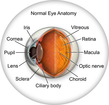
Don’t remember the lessons on eye anatomy from your high school biology class? That’s OK—we have provided the following eyeball illustration and terms just to give you a refresher course. And we won’t give you a pop quiz afterwards…
IRIS: Pigmented tissue lying behind cornea that (1) gives color to the eye, and
(2) controls amount of light entering the eye by varying size of black
pupillary opening; separates the anterior chamber from the posterior chamber.
black circle and it regulates the amount of light that enters the eye.
rays of light to focus on the retina.
CILIARY BODY: a muscular ring under the surface of the eyeball; helps the eye focus by changing the len’s shape and also produces aqueous humor
CHOROID: the vascular layer between the sclera and the retina; the blood vessels in the choroid help provide oxygen and nutrients to the eye
OPTIC NERVE: Largest sensory nerve of the eye; carries impulses for sight from
retina to brain.
MACULA: Small, specialized central area of the retina responsible for acute
central vision.
RETINA: Part of the eye that converts images into electrical impulses sent
along the optic nerve for transmission back to the brain. Consists of
many named layers that include rods and cones.
VITREOUS: Transparent, colorless, gelatinous mass; fills rear two-thirds
of the interior of the eyeball, between the lens and the retina.